Wu Liu1,
Guo-Shuai Feng1,2,
Yang Ou1,
Jun Xu3,
Zai-Jun Zhang1,
Gao-Xiao Zhang1,
Ye-Wei Sun1,
Sha Li4,
Jie Jiang1,5 ![]()
For correspondence:- Jie Jiang Email: jiangjie@jnu.edu.cn Tel:+862085222156
Received: 12 December 2015 Accepted: 2 July 2016 Published: 30 August 2016
Citation: Liu W, Feng G, Ou Y, Xu J, Zhang Z, Zhang G, et al. Neuroprotective effect of apocynin nitrone in oxygen glucose deprivation-treated SH-SY5Y cells and rats with ischemic stroke. Trop J Pharm Res 2016; 15(8):1681-1689 doi: 10.4314/tjpr.v15i8.13
© 2016 The authors.
This is an Open Access article that uses a funding model which does not charge readers or their institutions for access and distributed under the terms of the Creative Commons Attribution License (http://creativecommons.org/licenses/by/4.0) and the Budapest Open Access Initiative (http://www.budapestopenaccessinitiative.org/read), which permit unrestricted use, distribution, and reproduction in any medium, provided the original work is properly credited..
Purpose: To investigate the neuroprotective potential of apocynin nitrone (AN-1), a nitrone analogue of apocynin, in rat brain tissue as a novel candidate for ischemic stroke treatment.
Methods: In vitro neuroprotection of AN-1 was studied in SH-SY5Y cells treated with oxygen glucose deprivation (OGD). Cell viability was measured using 3-(4,5-dimethyl-2-thiazolyl)-2,5-diphenyl-2H-tetrazolium bromide (MTT) assay, and intracellular reactive oxygen species (ROS) level was investigated using flow cytometry. The protection of AN-1 in cerebral ischemia-reperfusion (I/R) rats was evaluated by cerebral infarct area and neurological deficit score. Oxidative stress of the ischemic hemisphere was assessed by malondialdehyde (MDA), glutathione (GSH) and superoxide dismutase (SOD) levels.
Results: In OGD-treated SH-SY5Y cells, AN-1 reduced cell death and ROS level. In I/R rats, AN-1 exerted potential protection against neurological deficit by reducing infarction area, decreasing neurological deficit score and relieving oxidative stress. AN-1 exhibited stronger action than its parent compound apocynin in vitro, but the two had similar effects in vivo. In addition, AN-1 demonstrated efficacy close to or higher than the positive reference Edaravone® both in vitro and in vivo. Furthermore, AN-1 showed lower toxicity than apocynin in vitro.
Conclusion: The results suggest that AN-1 may be a potential neuroprotective agent for the treatment of ischemic stroke in human.
Introduction
Ischemic stroke is a disturbance caused by occlusion of the cerebral vasculature, which lead to decreased delivery of glucose and oxygen to the brain, disruption of the ionic equilibrium and calcium homeostasis, excitotoxicity and eventual cell death [1,2]. It is one of the leading causes of death and disability worldwide. However, few therapeutic methods or drugs have adequate effects in the clinical treatment of ischemic stroke [2,3]. Timely recanalization of cerebral artery vessels and reintroduction of blood flow allow ischemic stroke-induced impairments to be arrested and reversed, thus, it is considered as an efficient therapy [3]. In ischemic cerebral injury, besides the ischemic infarction, the reperfusion after ischemia brings oxidative injury induced by reactive oxygen species (ROS) as well, which might cause delayed neuronal death [3]. To prevent irreversible ischemic injury, neuroprotection is essential [2-4]. In ischemia-reperfusion (I/R), superoxide anion is one of the most important ROS, as it contributes to oxidative injury in the progress of ischemic stroke [5]. NADPH oxidase (NOX) can effectively generate superoxide anion, which further interacts with nitric oxide to form peroxynitrite, another key reactive species accounting for neuronal death [6]. NOX is a major mediator involved in I/R-induced neuronal death. Thus, NOX inhibitors or genetic deletion of NOX have potential as treatments for ischemic injury due to stroke [7,8].
Apocynin (Apo, 4-hydroxy-3-methoxyacetophe-none), a natural compound in the root of Picrorhiza kurroa Royle, is able to specifically block the activity of NOX by preventing the transfer of its cytosolic subunit to membrane [7]. In previous researches, Apo was reported to improve ischemic infarction and show neuroprotection against middle cerebral artery occlusion (MCAO)-induced impairments in wild-type rodent models but not in NOX2-deficient mice [7,8]. However, Tang et al reported that the therapeutic dose range of Apo was quite narrow in experimental stroke [9]. Hence, new apocynin analogues were designed and synthesized in our lab to explore promising candidates in treating ischemic stroke [10].
Nitrones were originally developed as free radical trapping agents in free radical chemistry [11]. Recently, it was discovered that nitrones protected biological systems against oxidative stress induced impairments [11-14]. Nitrones have been tested as therapeutic agents for various diseases, including stroke [11,12]. In light of the neuroprotective activity of both apocynin and nitrones, we synthesized apocynin nitrone (AN-1) by conjugating apocynin with a nitrone moiety at the ortho position of its phenolic hydroxyl group [10].
In previous studies, AN-1 showed relatively good NADPH oxidase inhibition and activity against oxidative injury both in vitro and in vivo, compared with its parent apocynin [15,16]. In this work, AN-1 was further studied for its efficacy on ischemic stroke through neuroprotective investigation. To evaluate the efficacy of AN-1, human neuroblastoma cell line (SH-SY5Y) treated by oxygen glucose deprivation (OGD) followed by reoxygenation was used for test in vitro, and rats with cerebral I/R caused by middle cerebral artery occlusion (MCAO) were studied in vivo. For comparison, Edaravone® (Eda), a powerful free-radical scavenger often used in cerebral I/R injury studies [17,18], was used as a positive reference, and the parent Apo was included as a control compound.
Methods
Chemicals and reagents
3-(4,5-dimethylthiazol-2-yl)-2,5-diphenyltetrazolium bromide (MTT), dimethyl sulfoxide (DMSO), triphenyltetrazolium chloride (TTC) and 2',7'-dichlorofluorescein diacetate (DCFH-DA) were obtained from Sigma-Aldrich (St. Louis, MO, USA). Dulbecco's Modified Eagle Medium (DMEM), fetal blood serum (FBS) and penicillin-streptomycin solution (100×) were supplied by Gibco (Shanghai, China). Assay kits for glutathione (GSH), malondialdehyde (MDA) and superoxide dismutase (SOD) were provided by Nanjing Jiancheng Bioengineering Institute (Nanjing, China). All other reagents were purchased from Sigma-Aldrich unless stated otherwise.
Cell culture under normal and OGD condition
Under normal condition, the human neuroblastoma cell line SH-SY5Y (purchased from ATCC) was maintained in DMEM containing 10 % FBS and supplemented with 100 U/mL penicillin and 100 g/mL streptomycin in a humidified atmosphere containing 5 % CO2 at 37 ºC. Under OGD condition, the cells were incubated in glucose-free DMEM at 37 ºC in a tri-gas incubator with 95 % N2 and 5 % CO2.
Cell protection effect by MTT assay
The cell protection effect of the tested agents was measured by MTT assy. All compounds including AN-1 were dissolved in DMSO and freshly prepared before use (final culture concentration of DMSO < 0.1 %). SH-SY5Y cells were dispensed in 96-well plate at a density of 1 × 10⁵ cells per well. After 24 h incubation under normal condition (5 % CO2 at 37 ºC), cells were treated with the tested agents for 8 h under OGD condition, and the cells were continuously cultured for an additional 12 h under normal condition. A 10 µl aliquot of 0.5 % MTT solution was added to each well followed by 4 h of incubation. Then, the medium was discarded and 100 μl of DMSO was added to each well to dissolve the formazan. The optical density of each well was measured at 570 nm using a microplate reader (Spectra MAX 340, Molecular Devices Co, CA, USA). Cell viability was expressed as a percentage of the value of control cells.
Intracellular ROS assay
SH-SY5Y cells were seeded on 6-well plates at a density of 5 × 10⁵ cells/well, and OGD insult was generated as described above. After 1 h of OGD insult, cells were washed with phosphate-buffered saline (PBS) and incubated with 10 μM DCFH-DA for 20 min. The cells were collected by D-hanks buffer at a density of 1 × 10⁵ - 1 × 10⁶ cells/mL. The intracellular ROS concentration was measured using DCFH-DA fluorescent probe and a flow cytometer (Becton, Dickinson and Company, NJ, USA).
Animals and grouping
Male Sprague-Dawley (SD) rats (250 ± 10 g) were obtained from Medical Experimental Animal Center of Guangdong Province. The animals were housed at a constant temperature (24 ± 1 °C) in a humidity-controlled (60 ± 10 %) environment under a 12 h light/dark cycle, and given standard laboratory food and water ad libitum. All animals were kept 7 days for acclimation before the experiments. All animal studies were approved and conducted in accordance with the Guidelines of Laboratory Animal Ethics Committee of Jinan University (approval ref no. 20140408110751). All animal procedures used in this study were also conducted with reference to the Guide for the Care and Use of Laboratory Animals (8th edition) of the National Research Council (US) [19]. The rats were divided randomly into seven groups, as follows: control, I/R alone, I/R with apocynin (2 mg/kg), I/R with Eda (6 mg/kg), I/R with a low dose of AN-1 (1.5 mg/kg), I/R with a medium dose of AN-1 (3 mg/kg), and I/R with a high dose of AN-1 (6 mg/kg).
Animal surgical procedures
Rats were anesthetized with chloral hydrate (10 %), which provided a stable plane of anesthesia for the full duration of the experiment. The right common carotid artery was exposed, the external carotid artery (ECA) and internal carotid artery (ICA) were isolated. The ECA was dissected distally, a coated suture (4.0 monofilament nylon suture, Beijing Sunbio Biotech Co. Ltd, Beijing, China) was introduced into the ECA lumen, and then gently advanced into the ICA lumen to block middle cerebral artery (MCA) blood flow. Following this, the incision was closed and penicillin was given intraperitoneally to prevent inflammation. Two hours after occlusion, Apo, Eda and AN-1 were administered via the tail vein. Five minutes later, the suture was removed to restore blood flow. Sham-operated rats were manipulated similarly, except MCA was not occluded. Regional cerebral blood flow (rCBF) was monitored by laser-Doppler flowmeter (Periflux System 5000, Perimed Inc.) with a flexible probe over the skull. rCBF was measured before ischemia, during MCAO and after reperfusion. Animals that did not show rCBF reduction of at least 70 % and animals that died after ischemia induction were excluded. A successful animal model showed a significant rCBF reduction after MCAO and obvious rCBF restoration after removing the suture. Core body temperature was monitored with a rectal probe and maintained at 37 °C throughout the procedure. All surgical procedures were performed under an operating stereomicroscope.
Measurement of neurological deficit
Neurological deficit of all the rats was measured using the Longa method at 24 h after reperfusion [20]. A 5-point scoring system was used, as follows: 0, no detectable deficit; 1, inability to extend the contralateral forelimb; 2, mild circling to the contralateral side; 3, falling to the contralateral side; and 4, no spontaneous motor activity. Neurological assessment was performed by an investigator blinded to the experimental treatment. After neurological behavior testing, the animals were euthanized, and the brains were collected for measurement of the cerebral infarct area and anti-oxidative activity.
Measurement of the cerebral infarct area
The brain of each rat was quickly removed, chilled at -20 °C for 20 min, and then sliced into 2 mm coronal sections using a rat brain matrix (Harvard Apparatus, Holliston, MA, USA). Sections were incubated in a 1 % TTC solution at 37 °C for 30 min and fixed with 4 % formalin solution. The images of the sections were captured using a digital camera on a solid-colored background plate that was neither red nor white. The infarct area was measured using Image J software and calculated as follows in Eq. 1.
Infarct area (%) = {I-(R-L)}/L × 100…………. (1)
where I is the area of ischemic region, R the area of ischemic right hemisphere, and L the area of normal left hemisphere.
Evaluation of anti-oxidative activity
For each rat, the ischemic hemisphere was dissected out and homogenized in PBS buffer. The samples were then centrifuged at 10,000 × g for 15 min at 4 °C. The protein in the supernatant was quantified using a BCA protein assay kit. Malondialdehyde (MDA), glutathione (GSH) and superoxide dismutase (SOD) assay kits were used for measurement according to the manufacturer instructions, respectively.
Statistical analysis
Results from in vitro studies were carried out at least three times independently and were expressed as the mean ± standard error of the mean (SEM). Results from in vivo studies are shown as mean ± standard deviation. The data were analyzed using GraphPad Prism 5.0 (GraphPad, San Diego, CA, USA). One-way analysis of variance (ANOVA) and the Student’s t-test were used to evaluate the statistical differences. A value of p < 0.05 was considered statistically significant.
Results
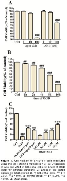
AN-1 protected SH-SY5Y cells against OGD-induced cell death
The cytotoxicity of AN-1 was first tested. The MTT results (A) indicated that under 10 μM, neither Apo nor AN-1 showed significant cytotoxicity in SH-SY5Y cells cultured under normal condition. However, both showed distinct cytotoxicity at 100 μM, and Apo was more toxic than AN-1. The test concentration of Apo and AN-1 was then controlled within 10 μM. As shown in B, OGD insult resulted in death of SH-SY5Y cells. Compared with the control group, the cell viability of OGD group dropped from 95.0 to 25.3 % after 1 to 16 h of OGD insult, respectively. 56.0 % of cell survived when treated with OGD for 8 h, thus, the OGD insult duration was set 8 h. C shows a low viability in OGD group, and demonstrates that Apo (10 μM) and Eda (1 μM) increased cell viability to 68.3 and 72.0 %, respectively. Meanwhile, AN-1 increased cell viability from 58.0 % to 79.0 % in a concentration-dependent manner.
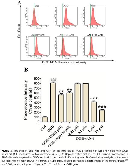
AN-1 reversed OGD-induced intracellular ROS increase
As shown in , compared with the control, OGD treatment (1 h) significantly induced ROS production in SH-SY5Y cells. Apo and Eda substantially decreased the level of ROS, while AN-1 reversed the production of ROS in a concentration-dependent way. The ROS production in the AN-1 group was markedly lower than that in the Eda and Apo groups at same concentration.
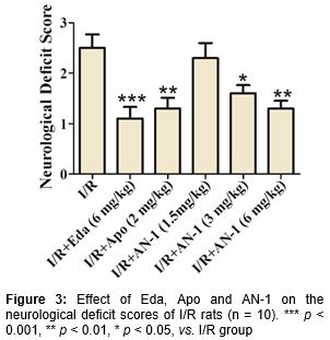
AN-1 ameliorated neurological deficit in I/R rats
The Longa method was adopted to assess the degree of neurological deficit of I/R rats after dosing of the tested agents. As shown in , rats in the I/R model group scored 2.5 after ischemia reperfusion. Apo and Eda decreased the scores to 1.3 and 1.1, respectively. AN-1 at 1.5 mg/kg played no role in ameliorating the neurological deficit, but reduced the neurological deficit scores to 1.6 and 1.3 at 3 and 6 mg/kg, respectively.
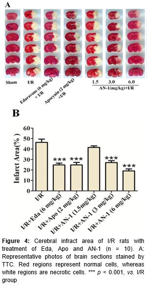
AN-1 reduced the MCAO-induced cerebral infarct area in rats
No infarct was observed in the brains of rats in the control group. However, large infract area was observed in the brains of I/R model rats. When treated with Apo (2 mg/kg), Eda (6 mg/kg) and AN-1 (3 and 6 mg/kg), the MCAO-induced cerebral infarct area in I/R rats was significantly reduced (p < 0.001). As shown in A and 4B, the cerebral infarct area (%) in I/R model rats was 46.4 %. Both Apo and Eda reduced the infarct area (%) to 24.8 %. AN-1 reduced the infarct area to 27.0 and 19.1 % at 3 and 6 mg/kg, respectively.
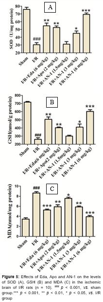
AN-1 reduced oxidative stress in I/R rats
Compared with the shame-operated control group (), the production of MDA increased by 151.6 %, and the level of SOD and GSH decreased by 59.9 and 63.7 %, respectively, in the brains of I/R model rats. Compared with the I/R model group, Eda and Apo significantly reversed the MDA increase (to 61.0 and 68.3 % of the I/R model group), SOD decrease (to 180.1 and 173.0 % of the I/R model group), and GSH decrease (to 194.9 and 155.1 % of the I/R model group). AN-1 reduced the production of MDA to 87.6, 68.0 and 45.6 % of the I/R model group at dose of 1.5, 3.0 and 6.0 mg/kg, respectively. Although AN-1 showed no distinct action to increase the level of SOD and GSH at 1.5 mg/kg, it enhanced the SOD (to 148.4 and 228.4 % of the I/R model group) and GSH (to 157.7 and 232.1 % of the I/R model group) levels at higher doses (3 and 6 mg/kg).
Discussion
Ischemic stroke results from blockage of blood supply to the brain. It brings irreversible injury of brain tissue if the cerebral blood vessel cannot be recanalized timely. Clinically, quick thrombolysis to restore blood flow as early as possible is recommended for the treatment of ischemic stroke [3]. The only proven thrombolytic therapy for acute ischemic stroke was achieved by intravenous use of t-PA. However, less than 10 % of patients with ischemic stroke may benefit from t-PA treatment because of the strict eligibility criteria for its use, including a relatively short therapeutic time window less than 4.5 h [21]. Follow-up care after blood flow restoration is also of great concern because I/R leads to serious brain injury [22,23]. Neuroprotective agents are thus urgently needed along with thrombolytic treatment to protect neurons from damage. Edaravone is an important agent, which was luached in Japan as a therapeutic for ischemic stroke through neuroprotection by free radical scavenging in 2001. However, some researchers have reported serious adverse reactions resulting from the use of Eda, including renal and hepatic disorders, this may partially account for its limited use [24,25]. While substantial numbers of experimental drugs have been investigated, so far, acute pharmacological intervention with neuroprotective agents has not been successful [2,4].
Oxidative stress is one of the prevalent factors in the development cascade of I/R injury from stroke [3,4,11,22,23]. As a NOX inhibitor, apocynin is a widely investigated anti-oxidative and anti-inflammatory agent [26]. Apocynin alleviated oxidative stress after I/R and demonstrated neuroprotective action in I/R MCAO mice by NOX inhibition [27]. Nitrone compounds, representing a group of free radical traps [11,12], demonstrated effective neuroprotection in animal stroke models [28,29]. In this work, AN-1 was synthesized by conjugating apocynin with nitrone to elicit potent neuroprotective action through oxidative stress amelioration.
In SH-SY5Y cells, AN-1 showed lower cytotoxicity than Apo at a high concentration, cell viability was almost 1.5-fold as much as that of Apo. At the same concentration, AN-1 was more powerful than Apo when it came to protect cells from OGD insult (p < 0.05). Moreover, the intracellular ROS production in the AN-1 group was 41.2 and 74.0 % of that in the Apo and Eda groups, indicating better antioxidant activity.
In MCAO model rats, AN-1 dose-dependently ameliorated neurological deficit induced by ischemia-reperfusion, although it exhibited no significant difference from the activity of Apo and Eda. The efficacy of AN-1 is similar to that of apocynin, while it is significantly greater than that of Eda in lessening cerebral infarct rate and palliating oxidative stress in the ischemic hemisphere (p < 0.05). The results in vitro and in vivo demonstrated that AN-1 has distinct anti-oxidative potency, which may be one of the chief mechanisms accounting for its neuroprotective action.
However, different researchers have reported widely different effective doses of Apo. Tang et al found that Apo improved neurological function (p < 0.01), and reduced the infarct volume (p < 0.05) and incidence of cerebral hemorrhage (p < 0.05) in mice when administered intravenously at a dose of 2.5 mg/kg but not at higher doses of 3.75 and 5 mg/kg, where it actually increased brain hemorrhage [9]. Interestingly, another group reported that Apo at 50 mg/kg attenuated cerebral infarction after transient focal ischemia in male SD rats [30]. In this study, Apo showed significant protective effect against MCAO-induced impairment at 2 mg/kg in male SD rats, while it easily triggered brain hemorrhage at 50 mg/kg in a trial test. Our results were inconsistent with those of Tang et al [30].
Conclusion
AN-1 enhances the cell survival rate and suppresses the over-production of ROS in OGD-treated SH-SY5Y cells. It protects ischemia-reperfusion rats from neurological deficit by alleviating cerebral infarction and oxidative stress, suggesting that AN-1 may be a potential neuroprotective candidate for ischemic stroke treatment.
Declarations
Acknowledgement
References
Archives


News Updates